
Dental X rays play a vital role in modern dentistry, serving as an essential diagnostic tool for detecting oral health issues before they become serious problems. While many patients express concerns about radiation exposure, understanding the facts about dental X rays can help alleviate these worries.
Today’s dental practices utilize advanced imaging technology and strict safety protocols to minimize radiation exposure while maximizing diagnostic benefits. This comprehensive guide explores the safety, purpose, and importance of dental X rays in maintaining optimal oral health.
What are Dental X Rays?
Dental X rays, also known as dental radiographs, are diagnostic imaging tools that use controlled bursts of radiation to create detailed images of teeth, gums, and surrounding bone structure. These images help dentists identify problems that aren’t visible during a regular visual examination, such as decay between teeth, bone loss from gum disease, or impacted wisdom teeth.
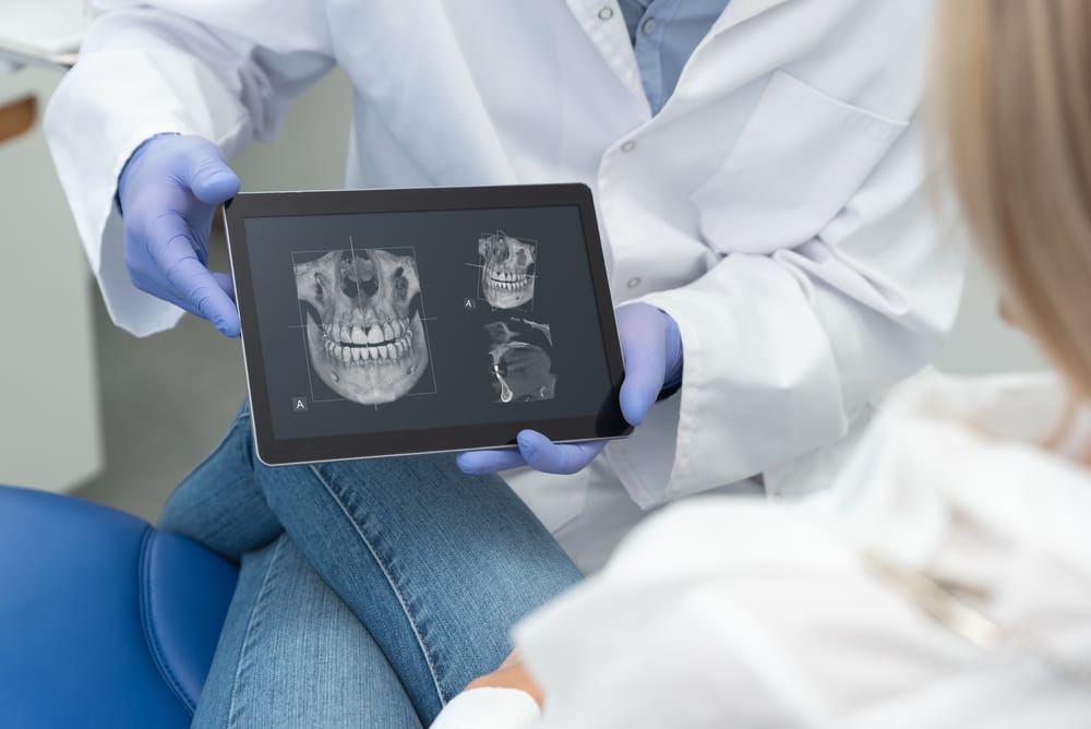
4 Types of Dental X Rays
- Bitewing X-rays: Focus on the crown portions of back teeth, showing areas where cavities often develop between teeth. These X-rays help detect early signs of decay and changes in bone density caused by gum disease.
- Periapical X-rays: Provide a view of the entire tooth from crown to root, including the surrounding bone. These images are essential for identifying deep decay, abscesses, cysts, and bone changes around a tooth’s root.
- Panoramic X-rays: Create a single image of all teeth and surrounding structures, including the jaw joints and sinuses. This type helps evaluate wisdom teeth, plan for implants, and check for abnormalities in the jaw.
- Occlusal X-rays: Show the floor or roof of the mouth, helping detect extra teeth, teeth that haven’t erupted, abscesses, or growths. These are particularly useful for examining children’s developing teeth.
What to expect during a Dental X-Ray?
- Preparation: The dental assistant will place a protective lead apron and thyroid collar on you to shield sensitive areas from radiation. You’ll also need to remove any metal objects like jewelry or dental appliances from your mouth.
- Positioning: The technician will position you in the dental chair, adjusting your head and bite position for optimal image capture. For some dental X rays, you might need to bite down on a small piece of plastic called a bitewing.
- Image Capture: The X-ray machine will be positioned around your head. The actual exposure only takes a fraction of a second, during which you’ll need to remain still to ensure clear images.
- Multiple Angles: Your dentist may need several images from different angles to get a complete view of the area being examined. The process will be repeated as necessary, with the technician repositioning the equipment each time.
- Review: After capturing all necessary images, the dentist will review them immediately on a computer screen (for digital X-rays) or after processing (for traditional film X-rays), then discuss any findings with you.
3 Key Benefits of Dental X Rays
Early Detection and Prevention
Regular dental X rays serve as a powerful preventive tool by identifying problems in their earliest stages. This early detection allows dentists to treat issues before they become painful or require more extensive, expensive procedures. The ability to spot potential problems early often means simpler, less invasive treatments and better long-term oral health outcomes.
Comprehensive Treatment Planning
Dental X rays provide dentists with crucial information needed to develop effective treatment plans. These detailed images help professionals map out complex procedures, determine the best approach for dental implants or orthodontic work, and track the progress of ongoing treatments. By having a clear view of the entire oral structure, dentists can make more informed decisions about treatment options.
Diagnosing Hidden Dental Issues
X-ray imaging reveals problems that remain invisible during routine visual dental examinations. Many serious dental conditions develop below the surface or in areas that cannot be seen directly, making dental X rays an invaluable diagnostic tool. Early identification of these hidden issues can prevent complications and reduce treatment costs.
- Tooth Decay Between Teeth: Cavities often develop in tight spaces between teeth where visual inspection and dental tools cannot reach. X-rays reveal these hidden decay spots before they cause significant damage.
- Advanced Gum Disease: Dental X rays show bone loss and deterioration beneath the gum line, helping identify periodontal disease progression that might not be evident during a visual exam.
- Impacted Teeth: These images reveal teeth trapped beneath the gum line or growing at awkward angles, particularly common with wisdom teeth.
- Bone Loss: X-rays detect decreasing bone density and structure changes that might indicate serious oral health issues or systemic diseases.
- Abscesses or Cysts: These infections at the root of teeth or in jawbones can be identified through X-ray imaging before they cause severe pain or spread to other areas.
Risks and Safety Considerations
Dental professionals carefully evaluate each patient’s needs before recommending X-rays, considering factors like age, oral health history, and current symptoms. They follow strict guidelines about the frequency and type of X-rays needed, ensuring that patients receive the minimum necessary exposure while still obtaining valuable diagnostic information. Modern dental practices prioritize patient safety through proper technique, equipment maintenance, and adherence to radiation safety protocols.
3 Contraindications
- Pregnancy: While dental X rays are generally safe, pregnant women should avoid routine X-rays during the first trimester unless absolutely necessary. Emergency X-rays can be performed with proper protection.
- Recent X-ray Exposure: Patients who have undergone multiple medical X-rays recently may need to postpone dental X rays unless urgently required.
- Thyroid Conditions: Patients with thyroid issues require additional protective measures and careful consideration of X-ray frequency.
4 General Risks of Dental X Rays
- Radiation Exposure: While dental X rays use very low radiation doses, repeated exposure should be monitored and limited to necessary diagnostic purposes.
- Equipment Malfunction: Improperly maintained or calibrated X-ray machines may deliver higher than necessary radiation doses, highlighting the importance of regular equipment maintenance.
- Operator Error: Incorrect positioning or multiple retakes due to poor technique can lead to unnecessary radiation exposure, emphasizing the need for properly trained professionals.
- Children’s Sensitivity: Young patients are more sensitive to radiation effects, requiring careful consideration of X-ray frequency and enhanced protective measures.
7 Guidelines and Regulations for Dental X Ray Safety
- Professional Training Requirements: All dental professionals who perform X-rays must complete specialized training and certification. This includes understanding radiation safety, proper equipment operation, and emergency protocols.
- Equipment Standards: Dental X-ray machines must meet strict FDA and state regulatory requirements. Regular inspections and calibration ensure equipment functions within safe operating parameters.
- Protective Equipment Usage: Dental offices must provide and properly maintain lead aprons and thyroid collars. These protective barriers should be inspected regularly for damage and replaced according to manufacturer guidelines.
- Image Quality Control: Dental practices must implement quality assurance programs to maintain optimal image quality while minimizing radiation exposure. This includes regular equipment testing and proper image processing procedures.
- Frequency Guidelines: Dental professionals must follow ADA recommendations for X-ray frequency based on patient age, risk factors, and oral health history. These guidelines help prevent unnecessary radiation exposure while maintaining diagnostic effectiveness.
- Patient Safety Protocols: Clear procedures must be established for identifying pregnant patients, documenting medical histories, and implementing appropriate safety measures. Special considerations apply for high-risk patients and children.
- Digital Technology Standards: Practices using digital X-ray systems must ensure proper data security, backup procedures, and software updates. Digital systems should be regularly calibrated to maintain accurate imaging while minimizing radiation exposure.
Conclusion
Dental X rays remain one of the most valuable diagnostic tools in modern general dentistry, combining minimal risk with maximum benefit when proper safety protocols are followed. Advanced technology, strict safety guidelines, and professional expertise ensure that patients receive the lowest possible radiation exposure while maintaining high-quality diagnostic imaging for optimal oral health care.
Visit Alki Dental to experience the latest in safe, digital X-ray technology and comprehensive dental care. Schedule your appointment today by calling our office or booking online. Stay connected with us on Facebook and Instagram for oral health tips and updates, and check out our reviews on Yelp to learn why our patients trust us with their dental care needs.
